|
基于miRNA测序分析清肺排毒汤防治小鼠急性肺损伤的分子机制研究
|
李龙雪 1(  ), 万崇凡 2, 张琦 2, 雷茹婷 2, 王潇玥 3, 程乐妍 2, 赖琦 2, 刘荣华 4( )(  ), 刘漩 1( )( ), 徐铁龙 5( )( )
|
|
Molecular Mechanisms of Qingfei Paidu Decoction in the Prevention and Treatment of Acute Lung Injury in Mice Based on miRNA Sequencing
|
LI Longxue 1( ), WAN Chongfan 2, ZHANG Qi 2, LEI Ruting 2, WANG Xiaoyue 3, CHENG Leyan 2, LAI Qi 2, LIU Ronghua 4( )( ), LIU Xuan 1( )( ), XU Tielong 5( )( )
|
|
|
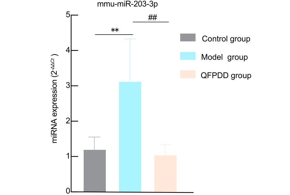
图 5. 实时荧光定量PCR验证mmu-miR-203-3p差异表达
注:对照组,连续8 d灌胃给予超纯水;模型组,灌胃给予超纯水1 d,然后于第2~8天给予脂多糖(LPS)水溶液雾化建立急性肺损伤模型;QFPDD组,连续8 d灌胃给予清肺排毒汤(QFPDD),并于第2~8天给予LPS水溶液雾化建模。每组随机选取4只小鼠的肺组织进行实时荧光定量PCR分析。与对照组相比,**P<0.01;与模型组相比,##P<0.01。
|
Figure 5. Validation of differential expression of mmu-miR-203-3p by quantitative real-time PCR
Note: Control group, intragastrically administered ultrapure water for 8 consecutive days. Model group, intragastrically administered ultrapure water on day 1, followed by exposure to aerosolized lipopolysaccharide (LPS) solution on days 2–8 to establish the acute lung injury (ALI) model. QFPDD group, intragastrically administered Qingfei Paidu decoction (QFPDD) for 8 consecutive days, with concurrent exposure to aerosolized LPS solution on days 2–8 for ALI induction. Lung tissues from 4 randomly selected mice per group were used for quantitative real-time PCR analysis (n=4). Compared with the control group, **P < 0.01; compared with the model group, ##P < 0.01.
|
|
 |
|
|