一种自研固定液用于制备大鼠眼球切片的组织学染色效果评价
|
|
唐小杭, 谷颖敏, 吕阳阳, 黄明姝, 田雪松
|
Evaluation of the Histological Staining Performance of Rat Eyeball Sections Prepared Using a Self-Developed Fixative
|
|
TANG Xiaohang, GU Yingmin, LÜ Yangyang, HUANG Mingshu, TIAN Xuesong
|
|
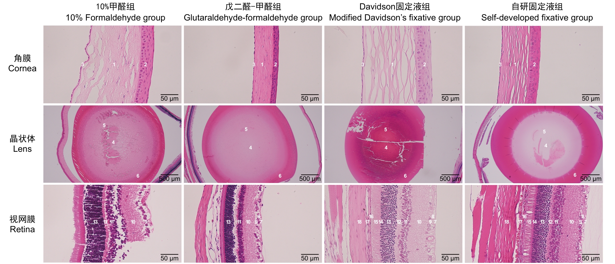
图2 显微镜下不同固定液组大鼠的眼球结构
注:显微镜下10%甲醛组、戊二醛-甲醛组、改良Davidson固定液组和自研固定液组的角膜、晶状体和视网膜HE染色结果。1,角膜基质层;2,角膜上皮层;3,角膜内皮层;4,赤道部;5,晶状体核区;6,晶状体皮质;7,内界膜;8,神经纤维层;9,节细胞层;10,内网层;11,内核层;12,外网层;13,外核层;14,外界膜;15,光感受器层;16,色素上皮层;17,脉络膜;18,巩膜。晶状体切片的比例尺为500 μm,角膜和视网膜切片的比例尺为50 μm。
|
Figure 2 Microscopic observation of rat eyeballs structures in different fixative groups
Note: HE staining results of the cornea, lens, and retina in the 10% formaldehyde group, glutaraldehyde-formaldehyde group, modified Davidson’s fixative group, and self?developed fixative group observed under light microscopy. 1, corneal stroma; 2, corneal epithelium; 3, corneal endothelium; 4, lens equator; 5, lens nucleus; 6, lens cortex; 7, inner limiting membrane; 8, nerve fiber layer; 9, ganglion cell layer; 10, inner plexiform layer; 11, inner nuclear layer; 12, outer plexiform layer; 13, outer nuclear layer; 14, external limiting membrane; 15, photoreceptor layer; 16, pigment epithelium layer; 17, choroid;18, sclera. Scale bar: 500 μm for lens sections; 50 μm for corneal and retinal sections.
|
|
|
|
|