外泌体通过NRF2/SLC7A11/GPX4通路调控铁死亡治疗小鼠缺血性脑卒中
|
|
徐英韬, 王蒙蒙, 林平, 迟海涛, 王怡, 白鹰
|
Exosomes Treat Ischemic Stroke by Regulation of Ferroptosis Through the NRF2/SLC7A11/GPX4 Pathway in Mice
|
|
XU Yingtao, WANG Mengmeng, LIN Ping, CHI Haitao, WANG Yi, BAI Ying
|
|
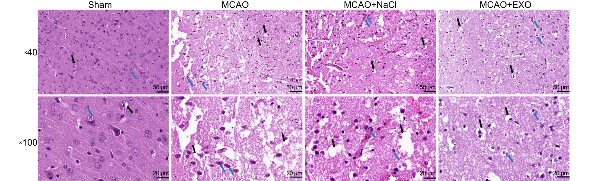
图4 各组小鼠脑组织切片HE染色观察神经细胞形态
注:假手术组(Sham)只暴露大脑中动脉,不电凝血管;模型组(MCAO)使用高频手术镊以8 W功率电凝大脑中动脉;模型+生理盐水组(MCAO+NaCl)先通过尾静脉注射100 μL生理盐水,然后进行电凝法造模;模型+外泌体组(MCAO+EXO)先通过尾静脉注射100 μL来自人羊膜间充质干细胞(hAMSCs)培养上清液的外泌体(9.5×10¹¹个/mL),然后进行电凝法造模。图中第1行×40视野比例尺为50 μm,第2行×100视野比例尺为20 μm。黑色箭头所指为细胞质空泡变性区域,蓝色箭头所指为细胞核固缩、碎裂区域。
|
Figure 4 HE staining of brain tissue sections from each group of mice to observe neuronal morphology
Note: The sham operation group (Sham) only exposed the middle cerebral artery without electrocoagulation of the vessel; the model group (MCAO) used high-frequency surgical forceps to electrocoagulate the middle cerebral artery at a power of 8 W; the model + normal saline group (MCAO+NaCl) first injected 100 μL of normal saline through the tail vein, and then performed electrocoagulation modeling; the model + exosome group (MCAO+EXO) first injected 100 μL of exosomes (9.5×10¹¹ particles per mL) from the culture supernatant of human amniotic mesenchymal stem cells (hAMSCs) through the tail vein, and then performed electrocoagulation modeling. The scale bar of the ×40 field of view in the first row of the figure is 50 μm, and the scale bar of the ×100 field of view in the second row is 20 μm. The area indicated by the black arrow is the region of cytoplasmic vacuolar degeneration, and the area indicated by the blue arrow is the region of nuclear pyknosis and fragmentation.
|
|
|
|
|