秀丽隐杆线虫的线粒体形态和功能研究方法及应用实例
|
|
宋梦娇, 沈义栋
|
Approaches and Application Examples for Studying Mitochondrial Morphology and Function in Caenorhabditis elegans
|
|
SONG Mengjiao, SHEN Yidong
|
|
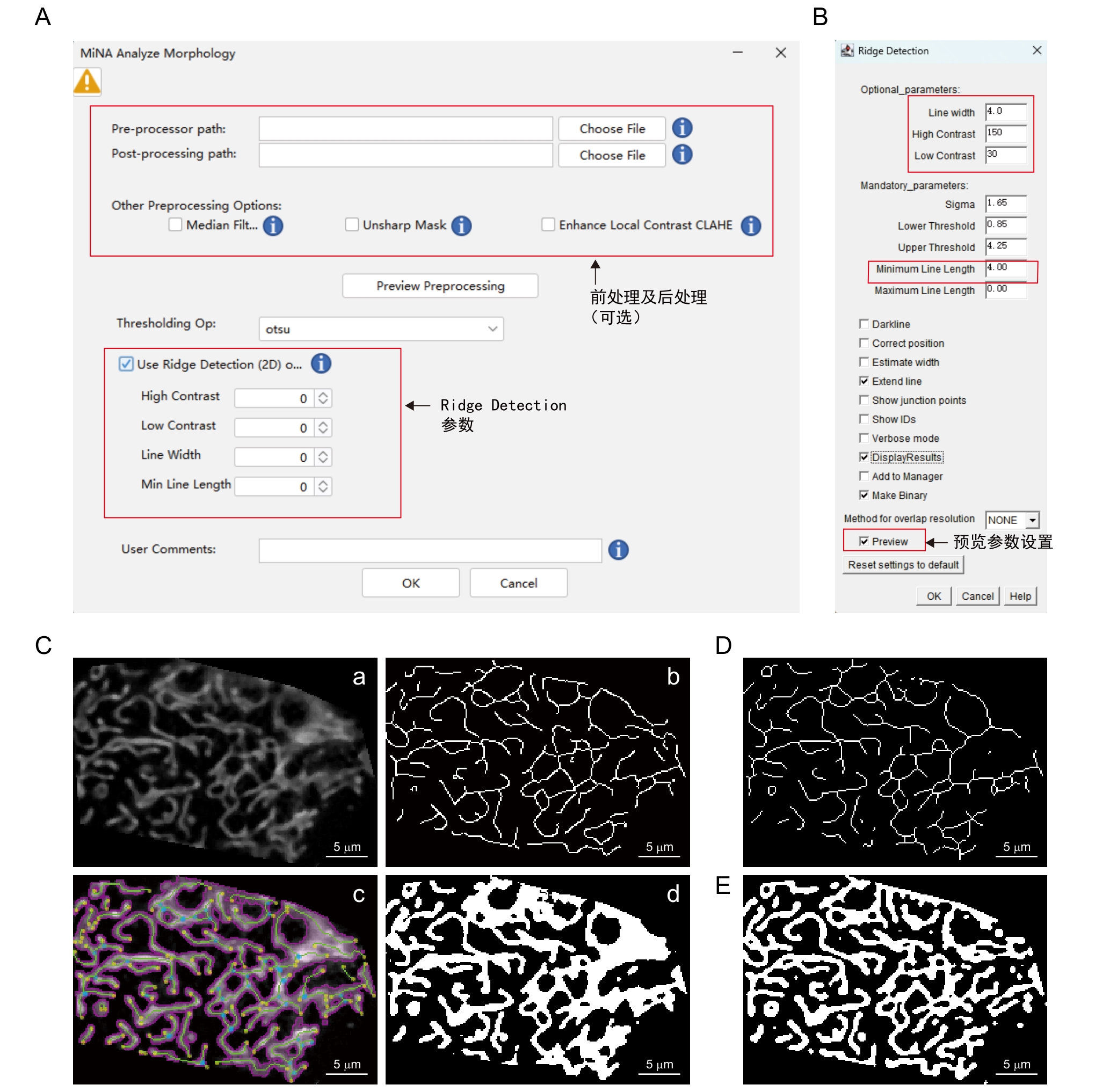
图5 MiNA - Mitochondrial Network Analysis插件步骤及注释
注:A~B,插件操作界面。C,软件处理模式图,将经过前处理的图像(a)通过自动阈值(Auto threshold-otsu)二值化图像获取线粒体轮廓(b),以Ridge Detection界面设定的参数(B)获取骨架图像(c),最后分别通过二值化图像和骨架图像计算相关参数并生成轮廓图 (d)。D,Skeletonize骨架算法生成结果。E,Mitochondria Analyzer二值化算法生成结果。
|
Figure 5 Procedures and annotations of MiNA - Mitochondrial Network Analysis plugin
Note: A-B, Plugin operation interface. C, Schematic diagram of the software processing workflow. The pre-processed image (a) undergoes binarization via the auto thresholding method (Auto threshold-Otsu) to extract mitochondrial contours (b). Using the parameters set for Ridge Detection in (B) to obtain the skeleton image (c). Relevant parameters are calculated from both the binarized image and skeleton image, and a contour map (d) is produced. D, Skeletonize algorithm(Skeletonization). E, Mitochondria Analyzer binarization algorithm.
|
|
|
|
|