秀丽隐杆线虫的线粒体形态和功能研究方法及应用实例
|
|
宋梦娇, 沈义栋
|
Approaches and Application Examples for Studying Mitochondrial Morphology and Function in Caenorhabditis elegans
|
|
SONG Mengjiao, SHEN Yidong
|
|
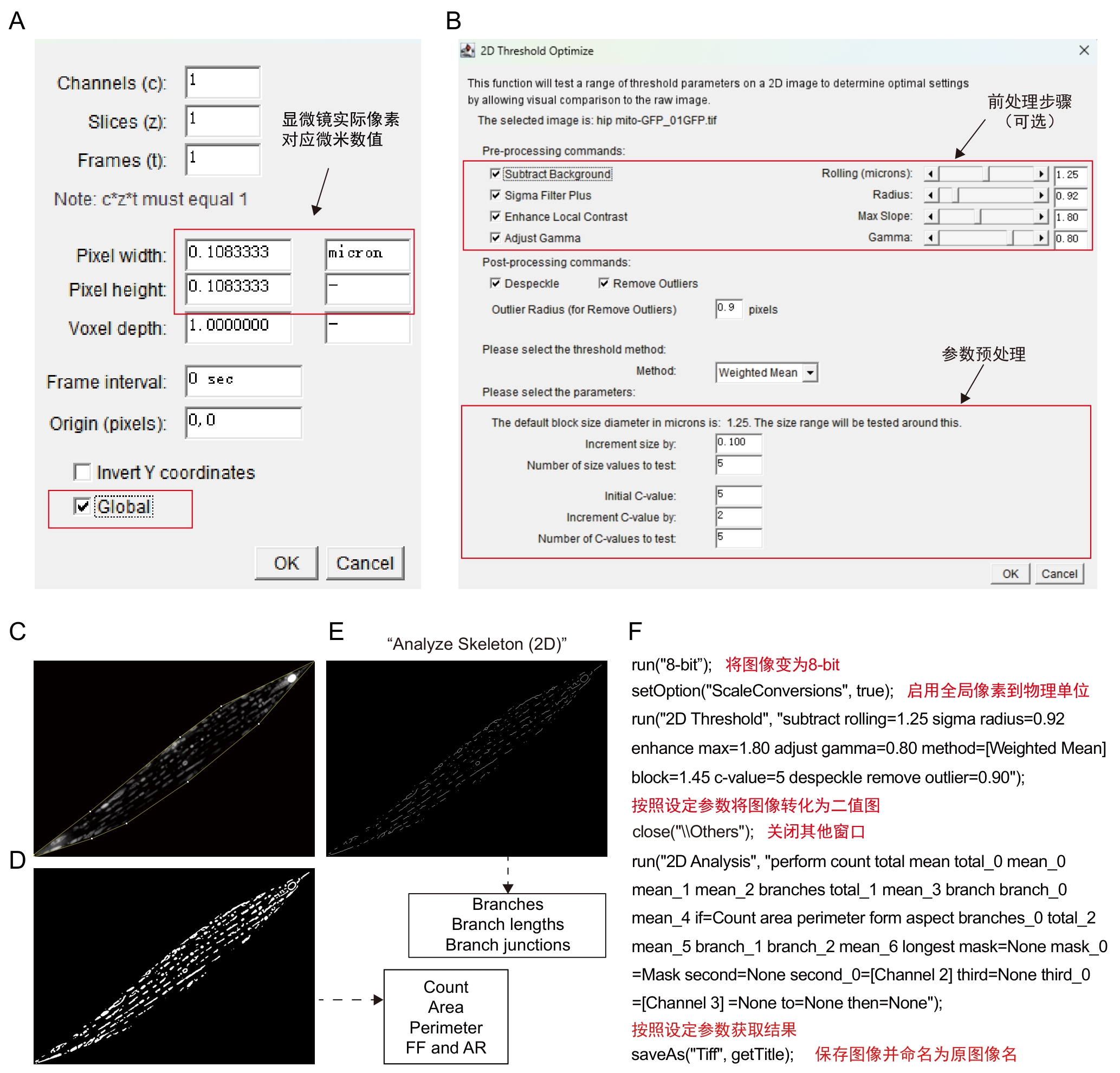
图4 Mitochondria Analyzer插件步骤及注释
注:A~B,插件操作界面。C~E,软件处理模式图,将经过前处理的图像(C)以设定的block size和C-value参数获取二值化图像(D),并在此基础上通过Skeletonize(2D)获取骨架图像(E)。二值化图像(C)可进一步获取线粒体数量(Count)、面积(Area)、周长 (Perimeter)、外形因数(form factor,FF)、长宽比(aspect ratio,AR)等数值,骨架图像(E)可进一步获取骨架分支数(Branches)、分支长度(Branch lengths)、骨架交汇点数(Branch junctions)等数值。F,C~E的批量处理宏及其注释。
|
Figure 4 Procedures and annotations of Mitochondria Analyzer plugin
Note: A-B, Plugin operation interface. C-E, Schematic of software processing workflow, the pre-processed image (C) is converted into a binarized image (D) using defined block sized and C-value parameters. The binarized image (D) is then skeletonized via the Skeletonize (2D) function to obtain the image (E) . From the binarized image (C), parameters such as mitochondrial count, area, perimeter, form factor and aspect ratio can be derived. From the skeleton image (E), data including branch number (Branches), branch lengths and branch junctions can be extracted. F, batch processing macro for steps C–E, accompanied by explanatory notes.
|
|
|
|
|