秀丽隐杆线虫的线粒体形态和功能研究方法及应用实例
Approaches and Application Examples for Studying Mitochondrial Morphology and Function in Caenorhabditis elegans
秀丽隐杆线虫的线粒体形态和功能研究方法及应用实例 |
| 宋梦娇, 沈义栋 |
|
Approaches and Application Examples for Studying Mitochondrial Morphology and Function in Caenorhabditis elegans |
| SONG Mengjiao, SHEN Yidong |
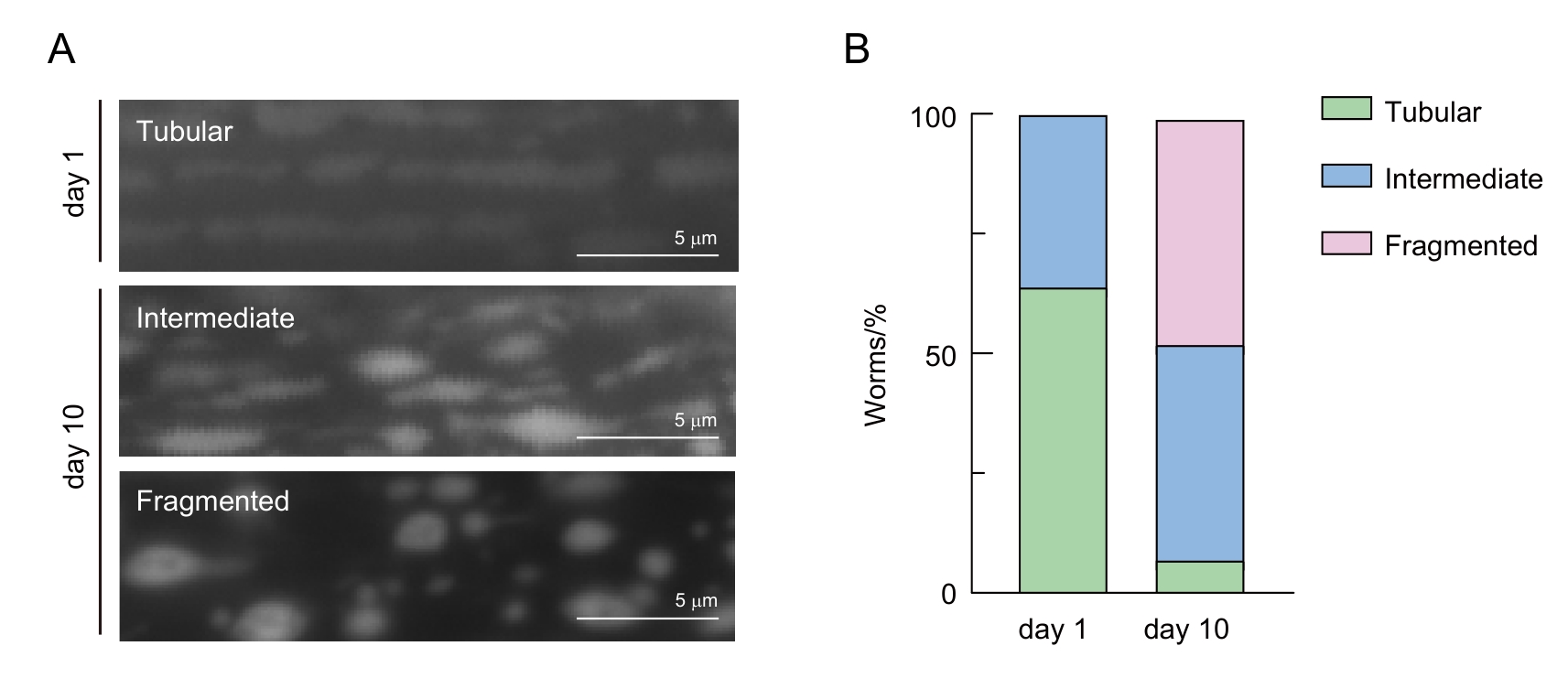
| 图3 线虫肌肉细胞线粒体形态分类 |
| Figure 3 Classification of mitochondrial morphology in C. elegans muscle cells |
|
|