大鼠胎儿期肺组织固定方法的探索
Exploration of Rat Fetal Lung Tissue Fixation Methods
大鼠胎儿期肺组织固定方法的探索 |
| 刘力瑜, 嵇波, 刘小玄, 方洋, 张玲, 郭亭廷, 全烨, 李鹤文, 刘翼天 |
|
Exploration of Rat Fetal Lung Tissue Fixation Methods |
| LIU Liyu, JI Bo, LIU Xiaoxuan, FANG Yang, ZHANG Ling, GUO Tingting, QUAN Ye, LI Hewen, LIU Yitian |
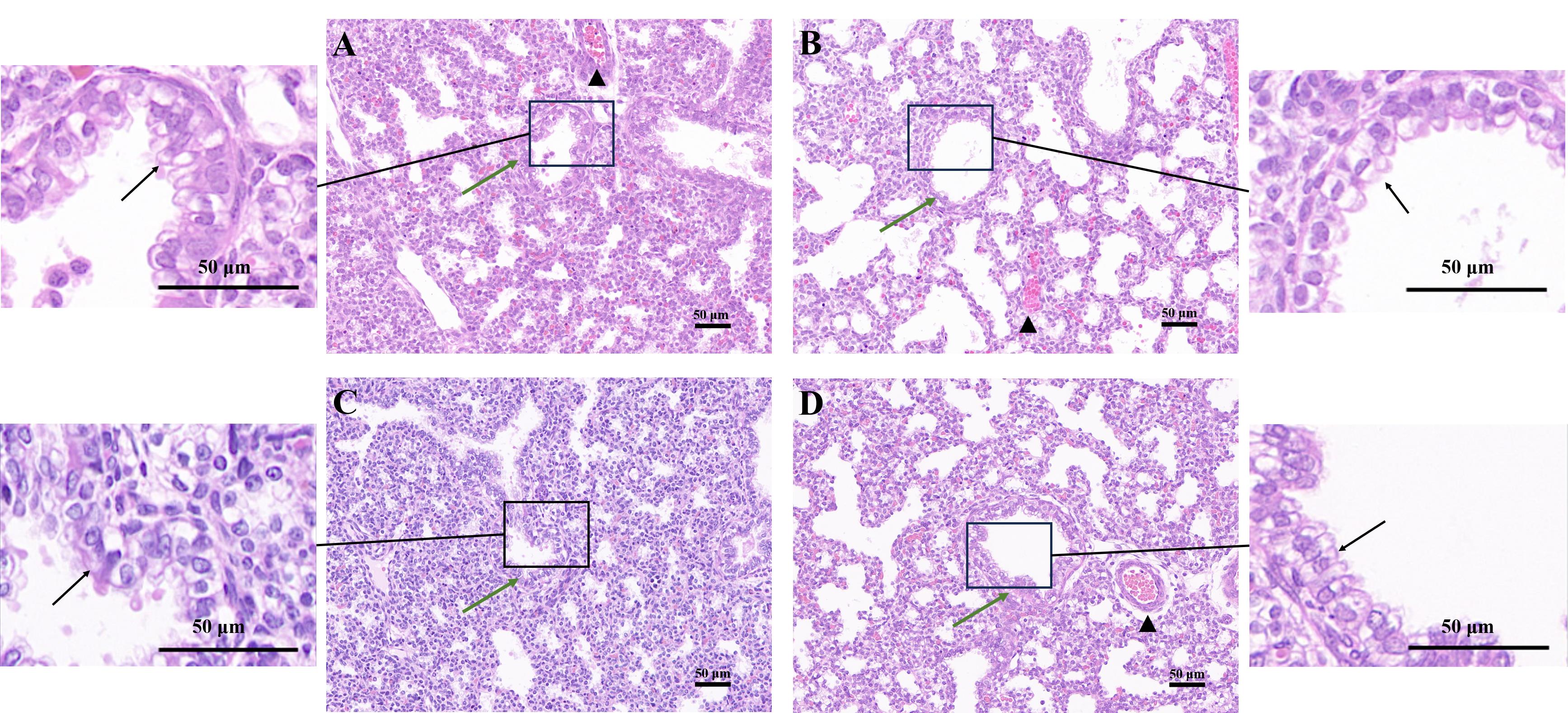
| 图4 四种固定方法下的胎鼠肺支气管HE染色结果(×200) |
| Figure 4 Results of HE staining of fetal rat lung bronchi usng four fixation methods (×200) |
|
|