大鼠胎儿期肺组织固定方法的探索
|
|
刘力瑜, 嵇波, 刘小玄, 方洋, 张玲, 郭亭廷, 全烨, 李鹤文, 刘翼天
|
Exploration of Rat Fetal Lung Tissue Fixation Methods
|
|
LIU Liyu, JI Bo, LIU Xiaoxuan, FANG Yang, ZHANG Ling, GUO Tingting, QUAN Ye, LI Hewen, LIU Yitian
|
|
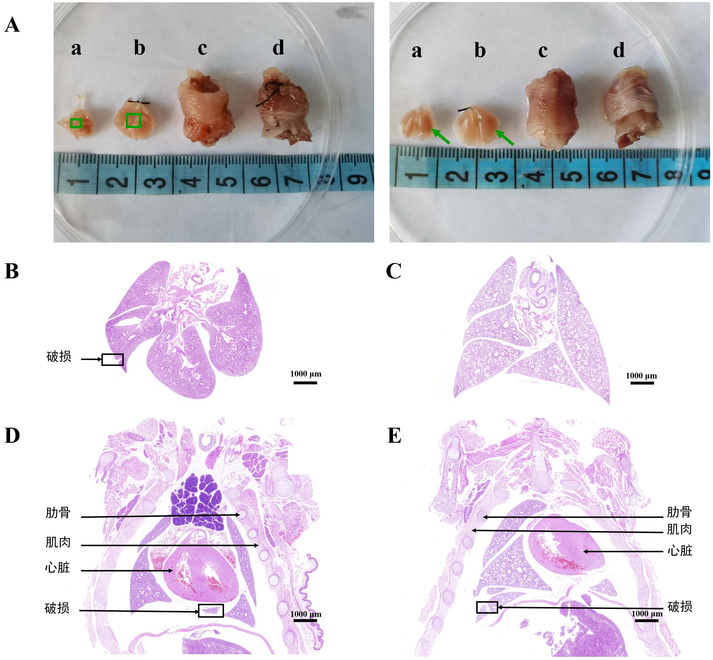
图2 四种固定方法下的胎鼠肺组织形态比较
注:A,胎鼠肺组织在四种固定方法下的大体观察结果,左图为冠状面仰卧位,右图为冠状面俯卧位;B~E为胎鼠肺组织在四种固定方法下的HE染色结果,其中B为直接取肺浸润法,C为气管内灌注后取肺浸润法,D为胎鼠整体浸润法,E为气管内灌注后胎鼠整体浸润法。绿色框内为肺门,绿色箭头所示为肺内血管,黑色箭头所示为肋骨、肌肉、心脏和肺脏破损处。
|
Figure 2 Comparison of fetal rat lung tissue morphology using four fixation methods
Note: A, comparison of fetal rat lung tissue morphology under gross observation using four fixation methods. The left image shows the coronal view in the supine position, while the right image displays the coronal view in the prone position. B-E show the HE staining results of fetal rat lung tissues under four fixation methods. B, direct lung infiltration method; C,lung infiltration method after intratracheal infusion; D, whole-body infiltration method of fetal rats; E, whole-body infiltration method after intratracheal infusion of fetal rats. The green boxes indicate the lung hilum, the green arrows indicate intrapulmonary blood and the black arrows indicate the ribs, muscles, heart, and sites of lung damage.
|
|
|
|
|