2019年某实验猴养殖场食蟹猴犬瘟热暴发的诊断
Diagnosis of an Outbreak of Canine Distemper in Cynomolgus Monkeys in an Experimental Monkey Farm in 2019
2019年某实验猴养殖场食蟹猴犬瘟热暴发的诊断 |
| 王晨娟, 杨玲焰, 王立鹏, 孙雪萍, 李静文, 郭连香, 荣荣, 时长军 |
|
Diagnosis of an Outbreak of Canine Distemper in Cynomolgus Monkeys in an Experimental Monkey Farm in 2019 |
| WANG Chenjuan, YANG Lingyan, WANG Lipeng, SUN Xueping, LI Jingwen, GUO Lianxiang, RONG Rong, SHI Changjun |
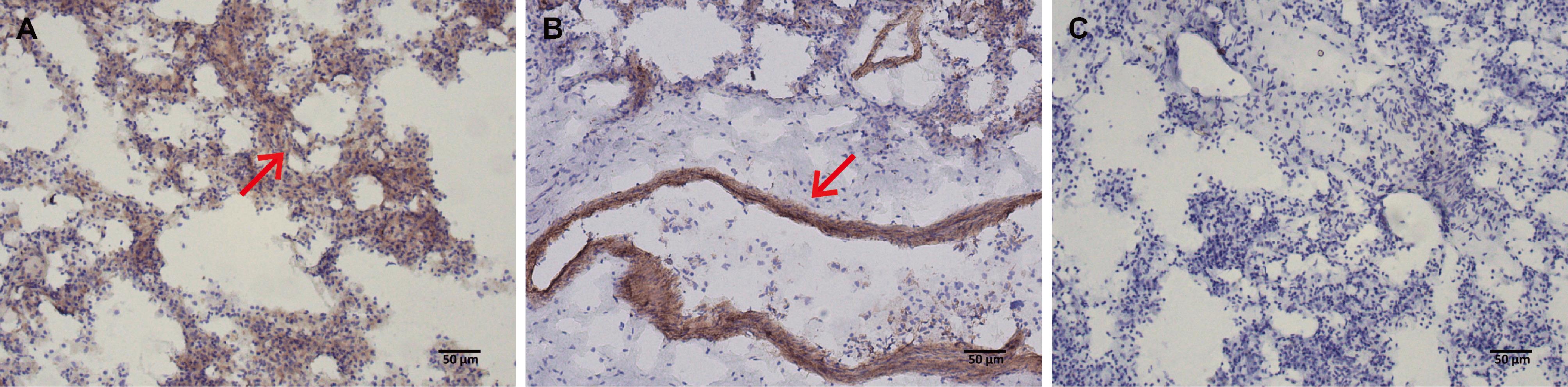
| 图2 免疫组织化学染色法检测病死猴肺组织切片中病毒蛋白表达(DAB染色,×100) |
| Figure 2 Detection of viral protein expression in lung tissue sections of the deceased monkey by immunohistochemical staining (DAB staining, ×100) |
|
|