利用脑立体定位技术将人源三突变APP基因导入海马区构建阿尔茨海默病大鼠模型
Establishment of a Rat Model of Alzheimer's Disease by Introducing Human Triple Mutant APP Gene into Hippocampus via Brain Stereotactic Technology
利用脑立体定位技术将人源三突变APP基因导入海马区构建阿尔茨海默病大鼠模型 |
| 肖林林, 杨逸萱, 黎珊杉, 罗兰诗雨, 尹思威, 孙俊铭, 施维, 欧阳轶强, 李习艺 |
|
Establishment of a Rat Model of Alzheimer's Disease by Introducing Human Triple Mutant APP Gene into Hippocampus via Brain Stereotactic Technology |
| XIAO Linlin, YANG Yixuan, LI Shanshan, LUO Lanshiyu, YIN Siwei, SUN Juming, SHI Wei, OUYANG Yiqiang, LI Xiyi |
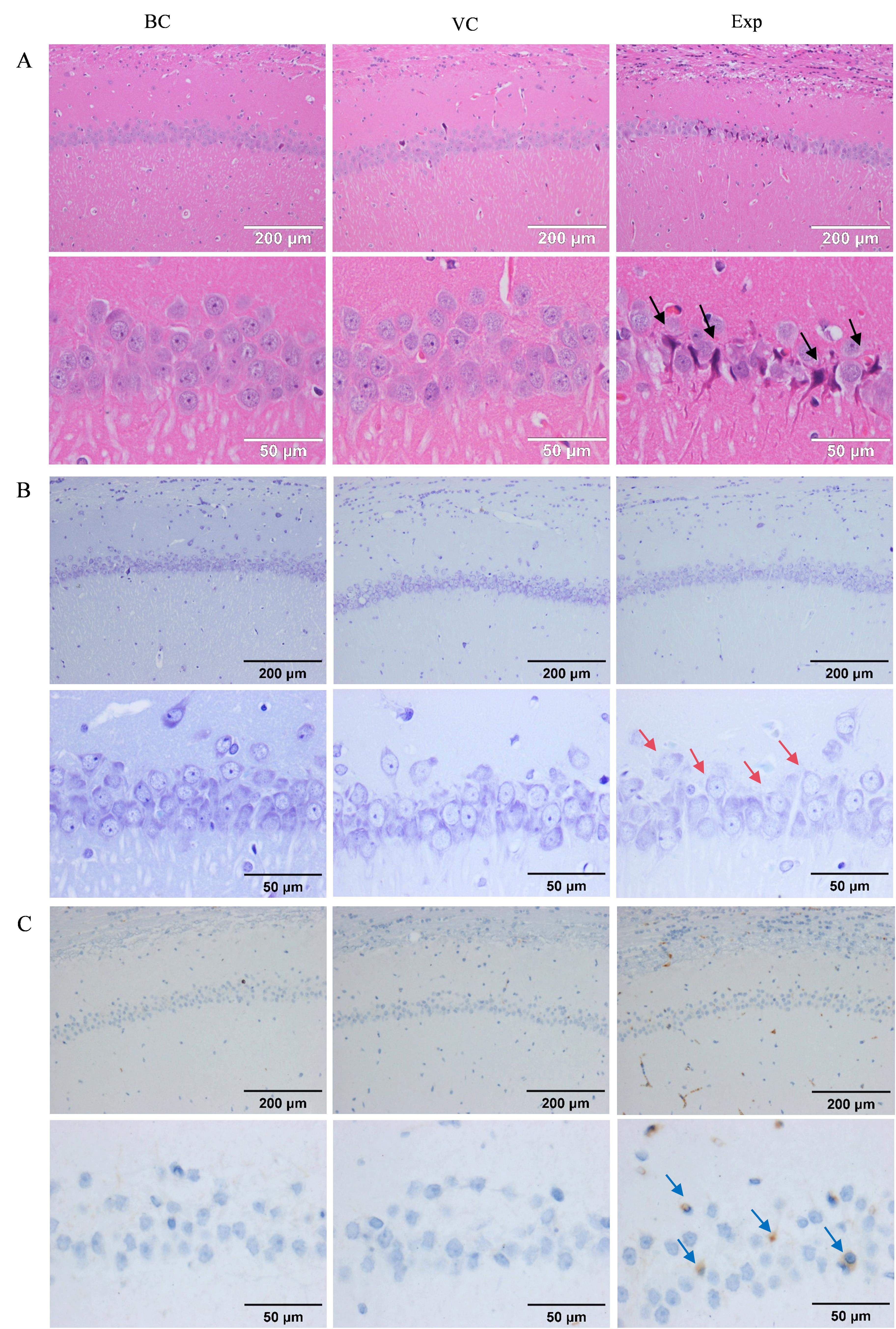
| 图5 大鼠海马体组织的HE染色(A)、尼氏染色(B)和免疫组织化学染色(C)结果 |
| Figure 5 HE staining (A), Nissl staining (B), and immunohistochemical staining (C) results of rat hippocampal region |
|
|