利用脑立体定位技术将人源三突变APP基因导入海马区构建阿尔茨海默病大鼠模型
|
|
肖林林, 杨逸萱, 黎珊杉, 罗兰诗雨, 尹思威, 孙俊铭, 施维, 欧阳轶强, 李习艺
|
Establishment of a Rat Model of Alzheimer's Disease by Introducing Human Triple Mutant APP Gene into Hippocampus via Brain Stereotactic Technology
|
|
XIAO Linlin, YANG Yixuan, LI Shanshan, LUO Lanshiyu, YIN Siwei, SUN Juming, SHI Wei, OUYANG Yiqiang, LI Xiyi
|
|
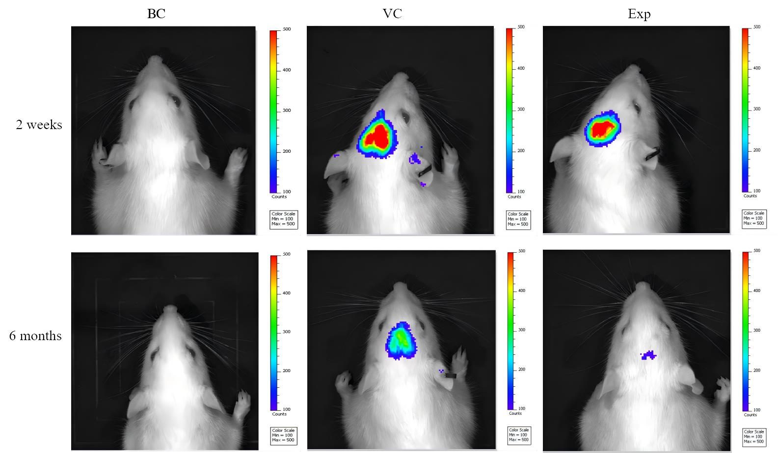
图2 注射病毒2周和6个月后大鼠活体成像情况
注:BC、VC、Exp分别为空白对照组、空载病毒组、实验组(通过脑立体定位在海马区注射携带人源三突变APP和NanoLuc萤光素酶基因的腺相关病毒)。各组大鼠腹腔注射萤光素酶底物Furimazine后10 min观察荧光素报告酶活性。2周后实验组与空载病毒组头部区域可检测到荧光发光,6个月后实验组和空载病毒组的荧光强度都减弱,但仍然可以被检测到;空白对照组未检测到荧光。
|
Figure 2 In vivo imaging of rats two weeks and six months after virus injection
Note:BC, VC, Exp are blank control group, virus control group, and experimental group (adeno-associated virus carrying human triple mutant APP and NanoLuc luciferase genes was injected in the hippocampus by brain stereotaxic localization), respectively. The luciferase reporter activity was observed 10 min after intraperitoneal injection of the luciferase substrate Furimazine into rats in each group. 2 weeks later, fluorescent luminescence could be detected in the head area of the experimental group and the virus control group, and 6 months later, the fluorescence intensity of both the experimental group and the virus control group was weakened, but it could be detected; the fluorescence was not detected in the blank control group.
|
|
|
|
|