长春瑞滨诱导大鼠足背静脉炎模型的动态评价
|
|
姜萌, 郝淑兰, 仝立国, 仲启明, 高振飞, 王永辉, 王晞星, 吉海杰
|
Dynamic Evaluation of Vinorelbine-Induced Phlebitis of Dorsalis Pedis Vein in a Rat Model
|
|
JIANG Meng, HAO Shulan, TONG Liguo, ZHONG Qiming, GAO Zhenfei, WANG Yonghui, WANG Xixing, JI Haijie
|
|
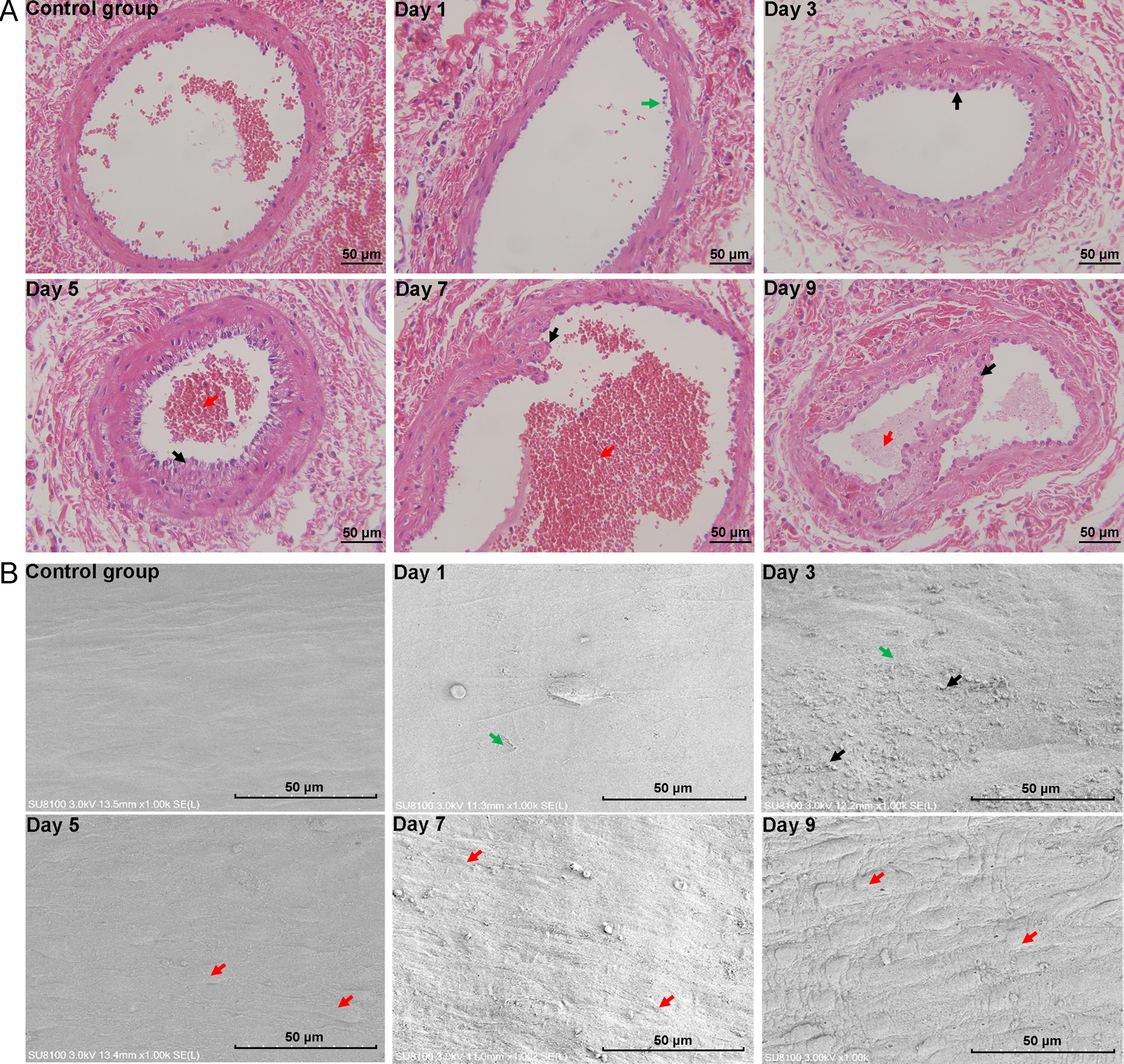
图2 足背静脉炎模型大鼠在不同时间点的静脉组织病理学(×400)和血管内膜微观结构(×1 000)
注:A为HE染色观察静脉组织病理学变化(绿色箭头指示内膜不规则破溃;黑色箭头指示内膜纤维性增生;红色箭头指示血栓);B为电子显微镜观察静脉血管内膜微观结构(绿色箭头指示内膜破溃;黑色箭头指示血细胞粘附;红色箭头指示内皮细胞隆起)。对照组大鼠于右后肢足背静脉注射0.1 mL生理盐水,模型组大鼠于右后肢足背静脉注射0.1 mL长春瑞滨溶液(4 mg/mL),连续观察9 d(Day 1、Day 3、Day 5、Day 7和Day 9分别为造模第1、3、5、7和9天)。
|
Figure 2 Histopathology of vein tissues (×400) and microstructure of vascular intima (×1 000) in phlebitis model rats at different time points
Note: A, histopathological changes of vein tissue stained by hematoxylin-eosin (HE) (green arrows indicate irregular rupture of the inner membrane; black arrows indicate intimal fibrous hyperplasia; red arrows indicate thrombosis); B, microstructure of vascular intima observed by scanning electron microscopy (SEM) (green arrows indicate inner membrane rupture; black arrows indicate adhesion of blood cells; red arrows indicate elevated endothelial cells). The control group rats were injected with 0.1 mL of saline solution into the dorsal vein of the right hind limb, while the model group rats were injected with 0.1 mL of vinorelbine solution (4 mg/mL). Observation continued for 9 consecutive days. Day 1, Day 3, Day 5, Day 7, and Day 9 refer to the first, third, fifth, seventh, and ninth days after modeling, respectively.
|
|
|
|
|