 ), 陆彩霞1()(
 )
), LU Caixia1()()
)
), LU Caixia1()()
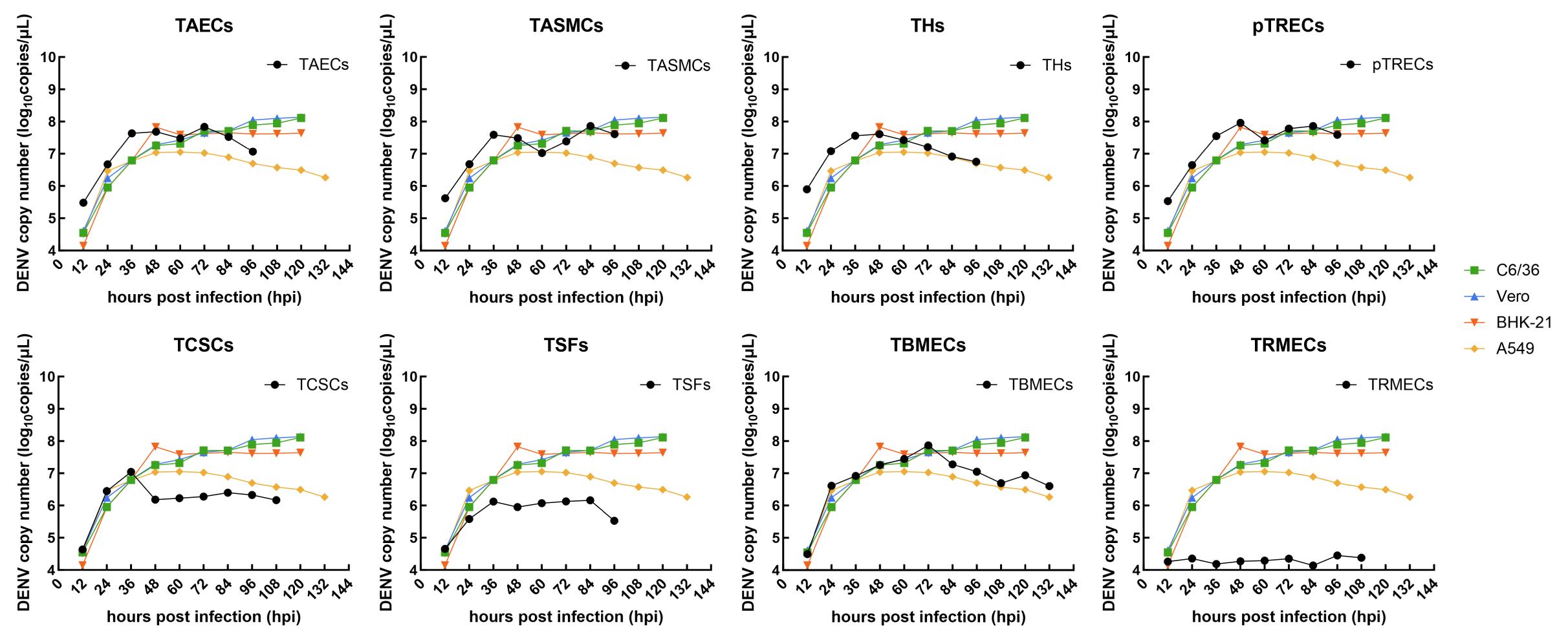
图2. 实时荧光定量PCR检测感染不同来源的树鼩细胞后登革病毒的增殖曲线
Figure 2. Viral proliferation curves of tree shrew cells of different tissues after dengue virus (DENV) infection via real-time fluorescent quatitative PCR
|
登革病毒对树鼩不同组织来源细胞的易感性及感染特性研究
|
|
刘欣1, 杞梦迪1, 王文广1, 罕园园1, 陆美丽1,2, 李娜1, 代解杰1(
), 陆彩霞1()()
|
|
Study on Susceptibility and Infection Characteristics of Dengue Virus in Cells Sourced from Different Tissues of Tree Shrews
|
|
LIU Xin1, QI Mengdi1, WANG Wenguang1, HAN Yuanyuan1, LU Meili1,2, LI Na1, DAI Jiejie1( ), LU Caixia1()()
|
|
图2. 实时荧光定量PCR检测感染不同来源的树鼩细胞后登革病毒的增殖曲线 |
Figure 2. Viral proliferation curves of tree shrew cells of different tissues after dengue virus (DENV) infection via real-time fluorescent quatitative PCR |
| |