|
登革病毒对树鼩不同组织来源细胞的易感性及感染特性研究
|
刘欣 1, 杞梦迪 1, 王文广 1, 罕园园 1, 陆美丽 1,2, 李娜 1, 代解杰 1(  ), 陆彩霞 1( )(  )
|
|
Study on Susceptibility and Infection Characteristics of Dengue Virus in Cells Sourced from Different Tissues of Tree Shrews
|
LIU Xin 1, QI Mengdi 1, WANG Wenguang 1, HAN Yuanyuan 1, LU Meili 1,2, LI Na 1, DAI Jiejie 1( ), LU Caixia 1( )( )
|
|
|
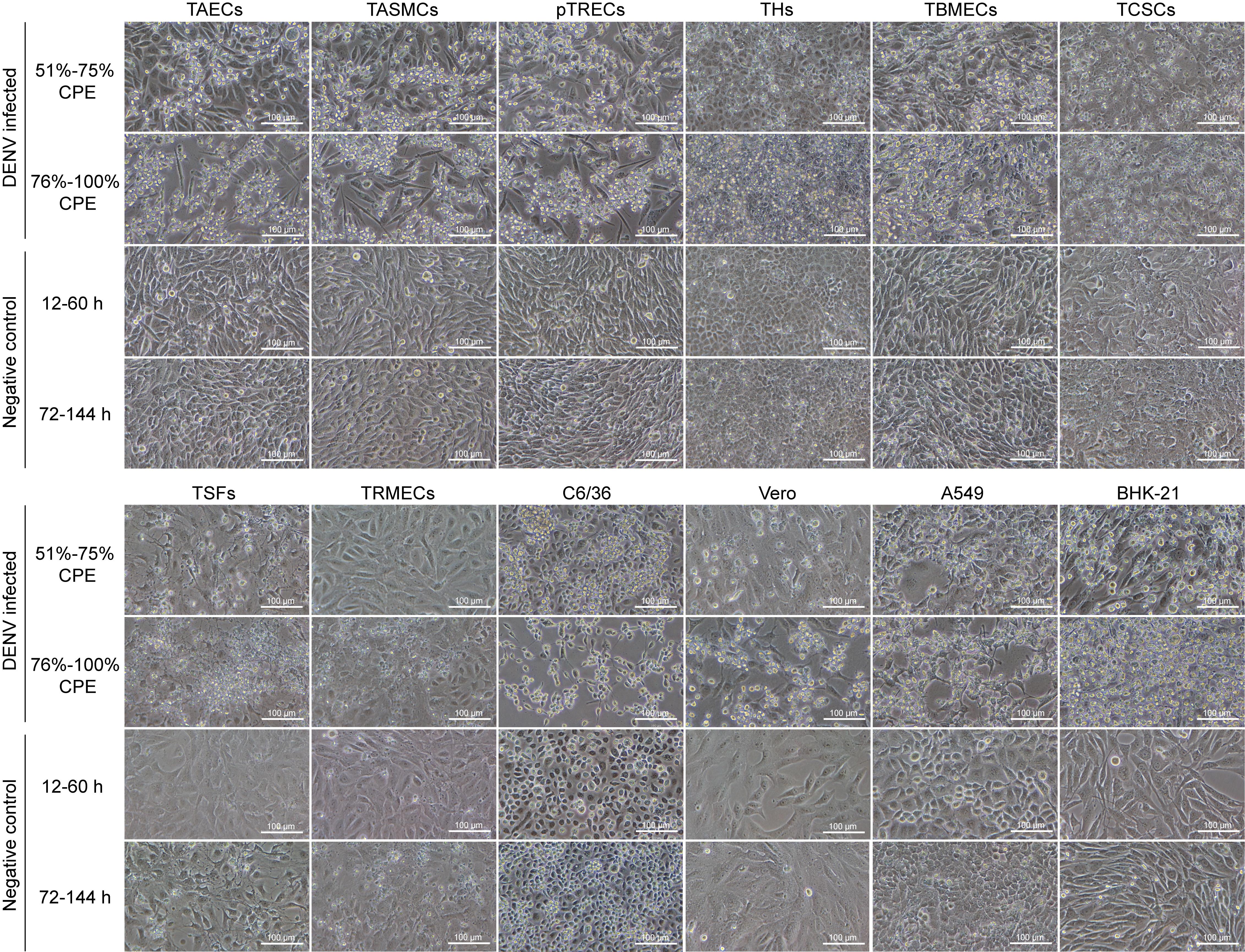
图1. 倒置显微镜下不同来源的树鼩细胞和阳性对照细胞感染登革病毒前后的形态学变化(×200)
注:TAECs为树鼩主动脉内皮细胞;TASMCs为树鼩主动脉平滑肌细胞;pTRECs为原代树鼩肾上皮细胞;THs为树鼩肝细胞;TBMECs为树鼩脑微血管内皮细胞;TCSCs为树鼩角膜基质细胞;TSFs为树鼩皮肤成纤维细胞;TRMECs为树鼩视网膜微血管内皮细胞;C6/36、Vero、A549及BHK-21作为阳性对照细胞。
|
Figure 1. Morphological changes in tree shrew cells of different tissues and positive control cells before and after dengue virus (DENV) infection under inverted microscope (×200)
Note: TAECs, tree shrew aortic endothelial cells; TASMCs, tree shrew aortic smooth muscle cells; pTRECs, primary tree shrew renal epithelial cells; THs, tree shrew hepatocytes; TBMECs, tree shrew brain microvascular endothelial cells; TCSCs, tree shrew corneal stromal cells; TSFs, tree shrew skin fibroblasts; TRMECs, tree shrew retinal microvascular endothelial cells; The C6/36, Vero, A549 and BHK-21 cells were employed as positive controls.
|
|
 |
|
|