异常子宫出血大鼠模型的构建与评价
连辉1, 姜艳玲1, 刘佳1, 张玉立2, 谢伟2, 薛晓鸥2, 李健1()()
Construction and Evaluation of a Rat Model of Abnormal Uterine Bleeding
LIAN Hui1, JIANG Yanling1, LIU Jia1, ZHANG Yuli2, XIE Wei2, XUE Xiaoou2, LI Jian1()()

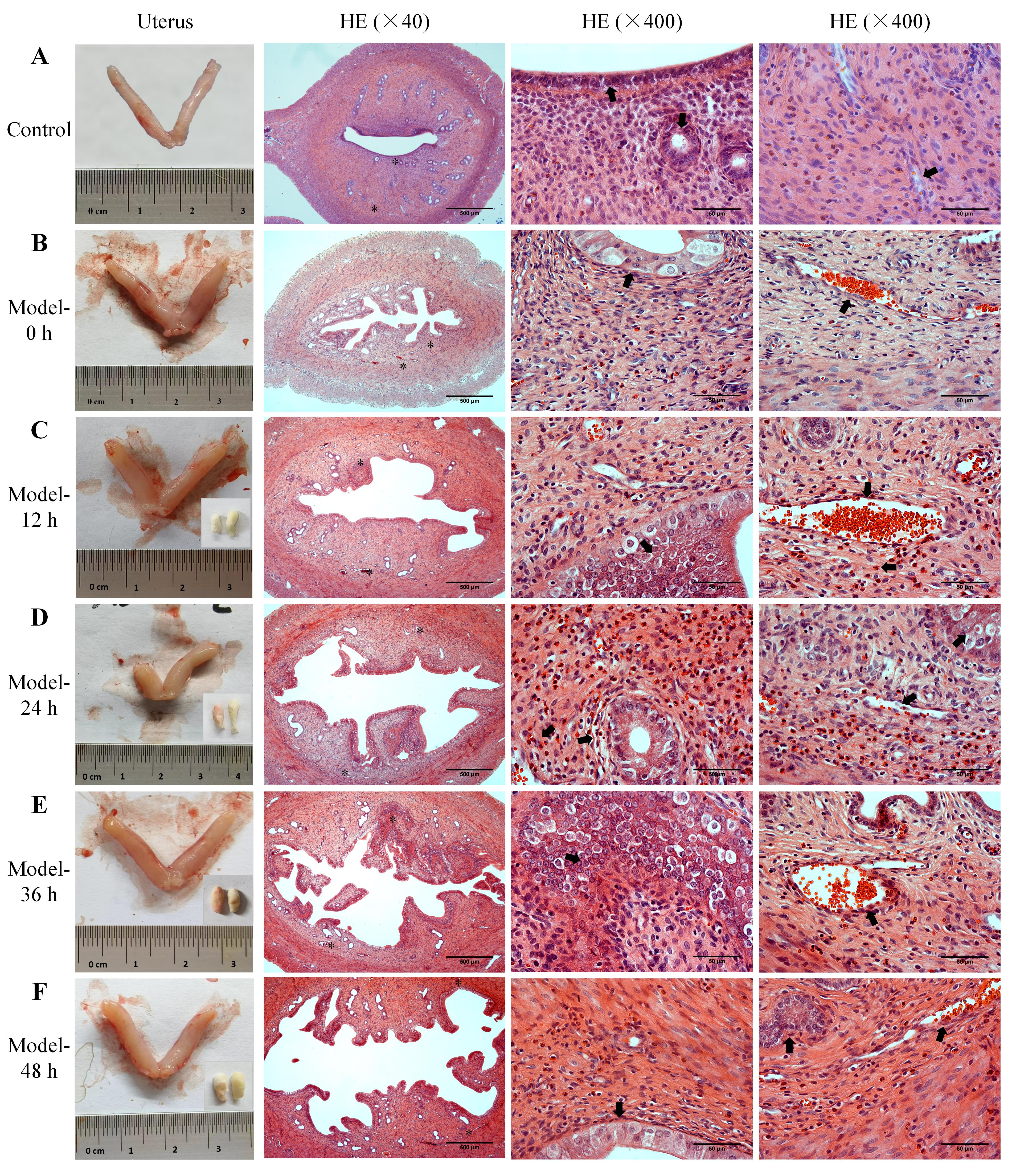
图3. 异常子宫出血模型大鼠的子宫外观、出血情况和内膜组织病理变化
注:A,正常对照大鼠的子宫外观和子宫内膜组织HE染色图片;B~F,异常子宫出血模型大鼠孕激素撤退0、12、24、36、48 h的子宫外观和收集每12 h子宫出血的阴道棉球(右下角)、子宫内膜组织HE染色图片。HE染色的低倍镜图片中两个星号(*)分别标记为两个高倍镜观察处,黑色箭头(?)指向内膜上皮、腺体、坏死的间质、血管和出血等结构和病理现象。HE染色图中比例尺从左到右分别为500 μm、50 μm和50 μm。

Figure 3. Uterine morphology, bleeding patterns, and histopathological changes of endometrial tissues in abnormal uterine bleeding model rats
Note: A, Uterine morphology and the HE-stained images of endometrial tissue in normal control rats; B-F, The uterine morphology in AUB model rats at 0 h, 12 h, 24 h, 36 h, and 48 h after progesterone withdrawal, along with vaginal cotton balls collected for uterine bleeding assessment every 12 hours (lower right corner), and the HE-stained images of endometrial tissue at the corresponding time points. The two asterisks (*) in each low-magnification HE-stained image indicate the two areas observed under high magnification, and the black arrows (?) point to the structures and pathological phenomena such as the endometrial epithelium, glands, necrotic stroma, blood vessels, and bleeding. In the HE-stained images, the scale bars from left to right column are 500 μm, 50 μm and 50 μm, respectively.