异常子宫出血大鼠模型的构建与评价
连辉1, 姜艳玲1, 刘佳1, 张玉立2, 谢伟2, 薛晓鸥2, 李健1()()
Construction and Evaluation of a Rat Model of Abnormal Uterine Bleeding
LIAN Hui1, JIANG Yanling1, LIU Jia1, ZHANG Yuli2, XIE Wei2, XUE Xiaoou2, LI Jian1()()

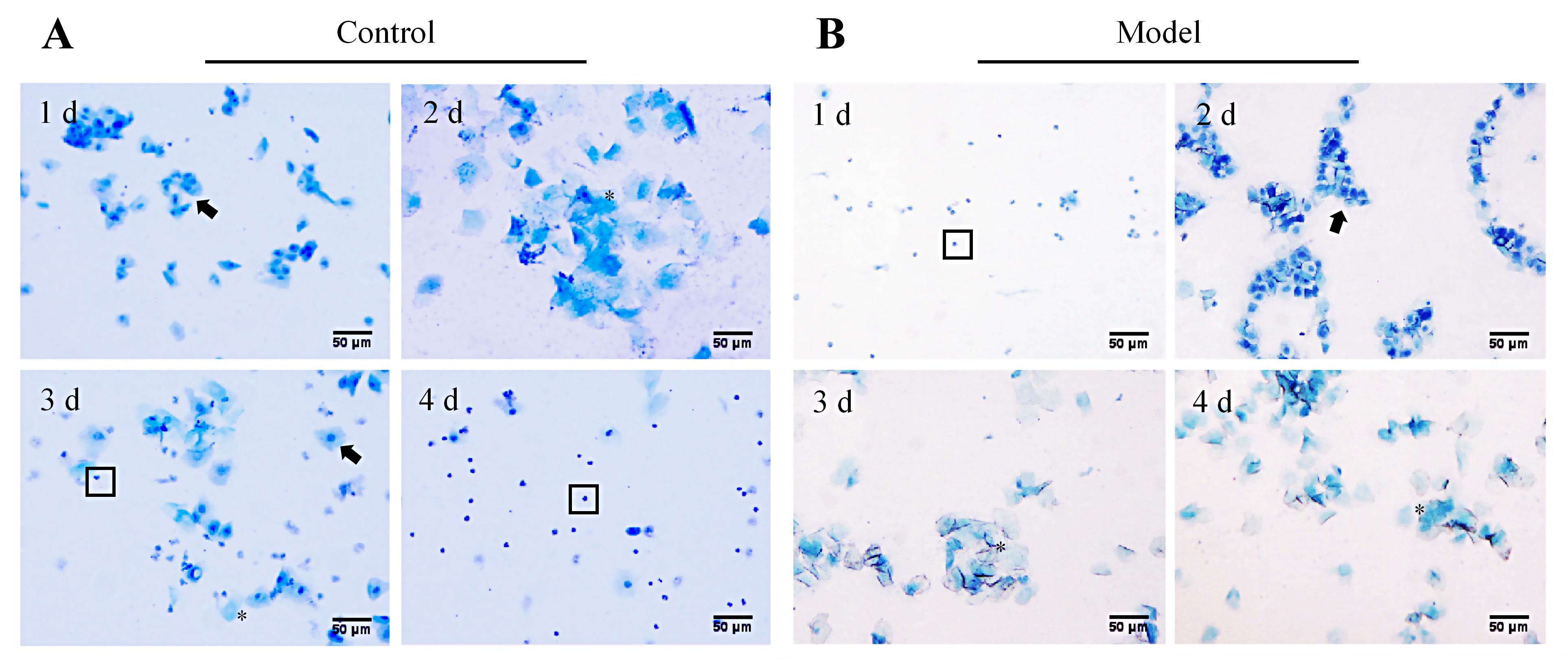
图 2. 异常子宫出血模型大鼠动情周期改变的阴道脱落细胞涂片检测(亚甲蓝染色,×200)
注:A,正常对照组一只代表性大鼠在一个动情周期内阴道脱落细胞的形态变化(1、2、3、4 d分别为动情前期、动情期、动情后期和动情间期,呈周期性改变);B,模型组一只代表性大鼠从卵巢切除、激素诱导到孕激素撤退的阴道脱落细胞形态变化(1、2、3、4 d分别为动情间期、动情前期、动情期和动情期,呈非周期性改变)。图片中黑色箭头(?)指向有核上皮细胞,星号(*)标记为角化上皮细胞,方框(□)内为白细胞。图中比例尺为50 μm。

Figure 2. Detection of changes in the estrous cycle in abnormal uterine bleeding model rats using vaginal exfoliated cell smear (methylene blue staining, ×200)
Note: A, Morphological changes of vaginal exfoliated cells of a representative rat in the normal control group during one estrous cycle (Days 1, 2, 3, and 4 correspond to proestrus, estrus, metestrus, and diestrus, respectively, showing cyclical changes); B, Morphological changes of vaginal exfoliated cells of a representative rat in the model group from ovariectomy, hormone induction to progesterone withdrawal (Days 1, 2, 3, and 4 correspond to diestrus, proestrus, estrus, and estrus, respectively, showing a non-cyclic pattern). In figures, black arrows (?) indicate nucleated epithelial cells, asterisks (*) mark keratinized epithelial cells, and boxes (□) denote white blood cells. In figures, the scale bar is 50 μm.