肝螺杆菌感染引起VDR-/-小鼠炎性肠病相关肠纤维化模型的建立及机制探讨
吴志浩1( ), 曹舒扬2, 周正宇1(
 )()
)()
), 曹舒扬2, 周正宇1()()
Establishment of an Intestinal Fibrosis Model Associated with Inflammatory Bowel Disease in VDR-/- Mice Induced by Helicobacter hepaticus Infection and Mechanism Exploration
WU Zhihao1(), CAO Shuyang2, ZHOU Zhengyu1()()
), CAO Shuyang2, ZHOU Zhengyu1()()
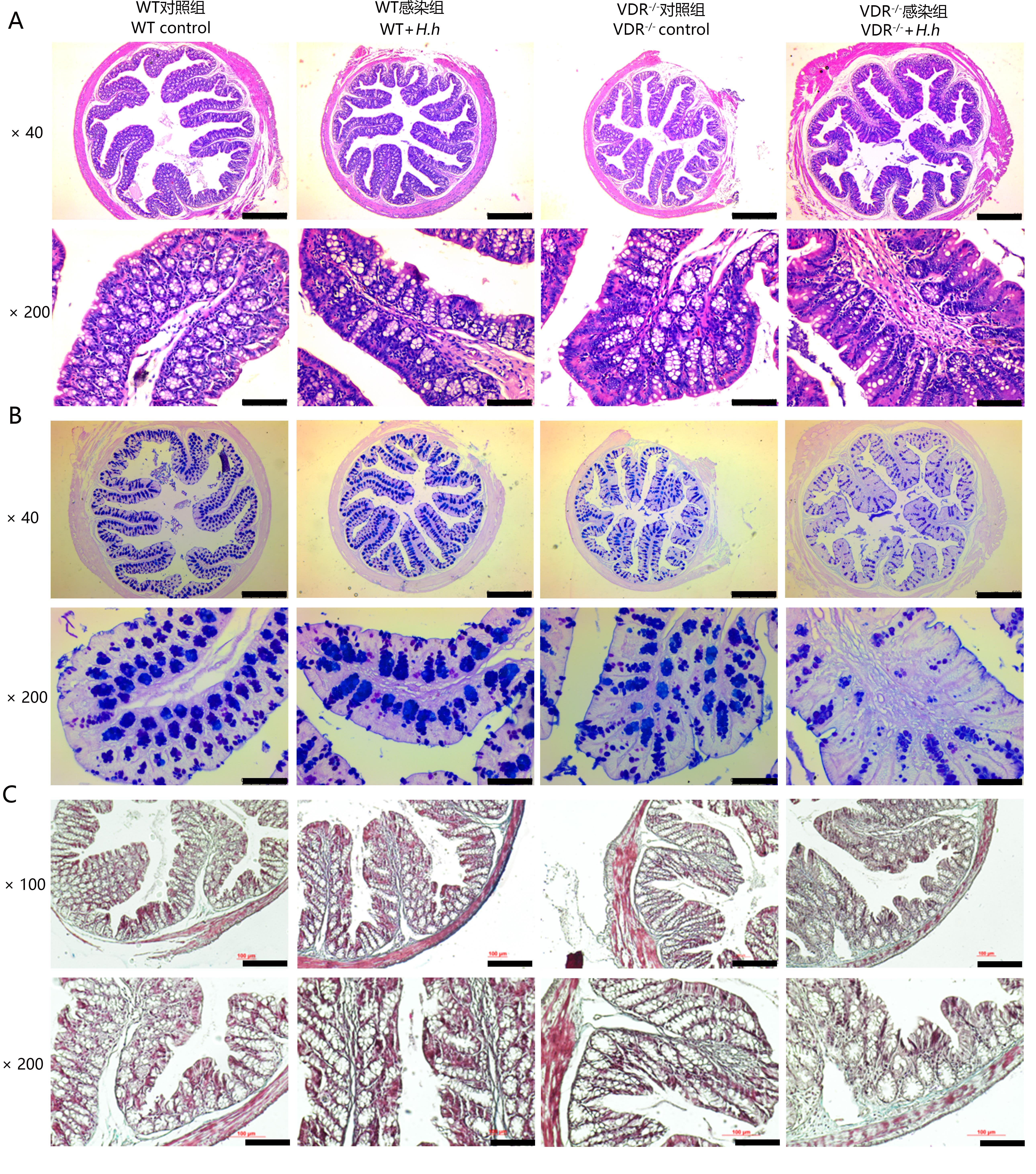
图4. H.hepaticus感染小鼠16周后结肠组织HE染色(A)、阿尔辛蓝-过碘酸希夫染色(B)和Masson染色(C)
Figure 4. HE staining (A), AB-PAS staining (B) and Masson staining (C) of colon tissue of mice infected with H.hepaticus for 16 weeks