|
SHJH hr 小鼠的心脏衰老表型研究
|
刘荣乐 1, 程灏 1, 尚付生 2, 常书福 1(  )(  ), 徐平 3
|
|
Study on Cardiac Aging Phenotypes of SHJH hr Mice
|
LIU Rongle 1, CHENG Hao 1, SHANG Fusheng 2, CHANG Shufu 1(  )(  ), XU Ping 3
|
|
|
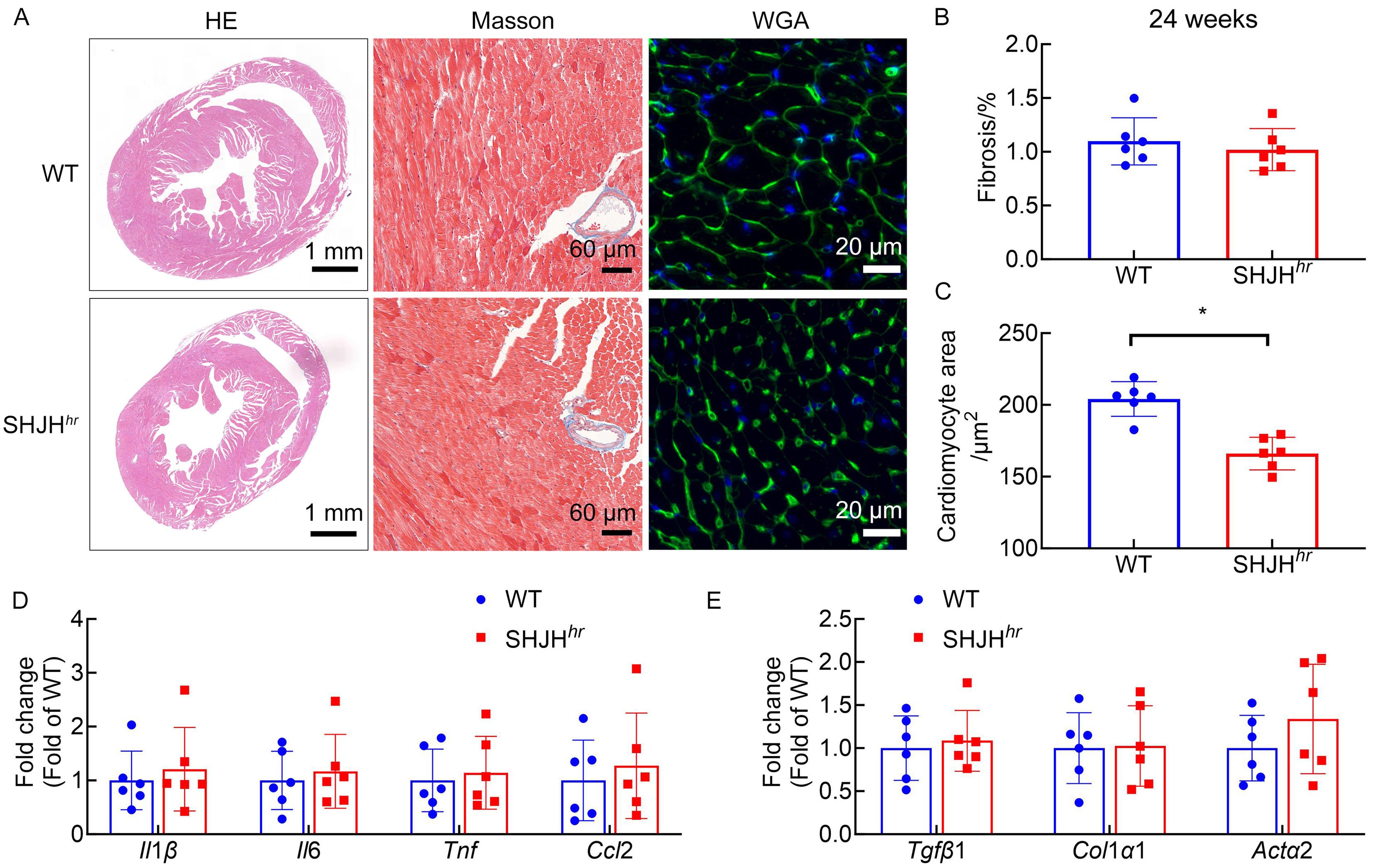
图3. 24周龄SHJH hr 小鼠的心脏组织病理和相关因子转录水平分析 注:A,HE、Masson 和WGA染色的代表性图片;B,Masson染色显示心肌纤维化水平统计;C,WGA染色显示心肌WGA表达细胞所占面积分析;D,实时荧光定量PCR(RFQ-PCR)检测心肌细胞中Il1β、Il6、Tnf和Ccl2基因的mRNA表达水平;E,RFQ-PCR检测心肌细胞中Tgfβ1、Col1α1和Actα2的mRNA表达水平。以非早衰的野生型ICR小鼠(WT组)作为对照,n=6;与WT组比较,*P <0.05。
|
Figure 3. Analysis of cardiac histopathology and related factor transcriptional levels in 24-week-old SHJH hr mice Note: A, Representative images of HE, Masson, and WGA staining; B, Statistical analysis of myocardial fibrosis levels shown by Masson staining; C, Analysis of the area occupied by WGA-positive cells in myocardial tissue as shown by WGA staining; D, Real-time fluorescent quantitative PCR (RFQ-PCR) measurement of mRNA expression levels of Il1β, Il6, Tnf, and Ccl2 in cardiomyocytes; E, RFQ-PCR measurement of mRNA expression levels of Tgfβ1, Col1α1, and Actα2 in cardiomyocytes. Wild-type ICR mice without premature aging (WT group) were used as controls, n=6. Compared with the WT group, *P <0.05.
|
|
 |
|
|