实验动物与比较医学 ›› 2021, Vol. 41 ›› Issue (3): 215-219.DOI: 10.12300/j.issn.1674-5817.2020.156

• 论著:人类疾病动物模型 • 上一篇    下一篇

松果体损伤大鼠模型3种制备方法的比较研究

王丹妮, 宋美卿, 杨钤, 冯玛莉   

  1. 山西省中医药研究院中心实验室,太原 030012
  • 收稿日期:2020-09-30 修回日期:2020-12-24 出版日期:2021-06-25 发布日期:2021-07-05
  • 作者简介:王丹妮(1993—), 女, 硕士研究生, 研究方向: 中药药理与内分泌疾病研究。E-mail: wdn0125@163.com
  • 基金资助:
    国家自然科学基金(81173574); 山西省自然科学基金(201901D111427)

Comparative Study of Three Pineal Gland Injury Model in Rats

WANG Danni, SONG Meiqing, YANG Qian, FENG Mali   

  1. Central Laboratory of Shanxi Academy of Traditional Chinese Medicine, Taiyuan 030012, China
  • Received:2020-09-30 Revised:2020-12-24 Online:2021-06-25 Published:2021-07-05

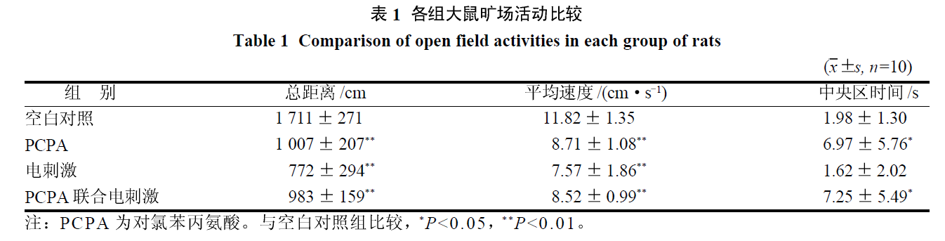
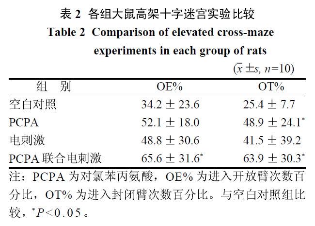
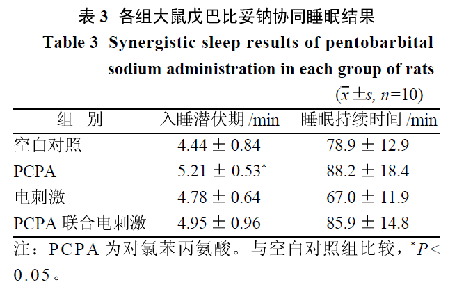
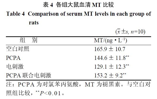
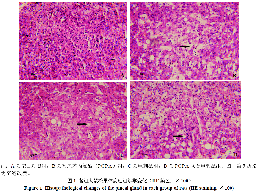
摘要: 目的 比较对氯苯丙氨酸(para-chlorophenylalanine,PCPA)、电刺激应激及PCPA联合电刺激3种不同方法制备的松果体损伤模型,评价松果体结构与功能变化,为建立方法简便、成模时间短的松果体损伤模型提供依据。方法 40只大鼠适应性饲养1周后,随机分为空白对照组、PCPA组、电刺激组及PCPA联合电刺激组。空白对照组不做任何处理;PCPA组大鼠腹腔注射PCPA 450 mg/kg,连续2 d;电刺激组大鼠使用穿梭箱电刺激(电压30 V,电流0.8 A,刺激时间30 s,间隔时间30 s,循环60次),连续5 d;PCPA联合电刺激组大鼠使用电刺激第4天加用PCPA,操作同前。测试各组大鼠旷场活动、高架十字迷宫等行为学以及戊巴比妥钠协同睡眠等指标,ELISA法检测血清褪黑素(melatonin,MT)水平,镜下观察松果体病理组织结构。结果 3个实验组大鼠旷场活动总距离和平均速度均显著低于空白对照组(P<0.01),PCPA组、PCPA联合电刺激组旷场活动中央区时间显著长于空白对照组(P<0.05),PCPA联合电刺激组进入开放臂次数显著多于空白对照组(P<0.05),PCPA组、PCPA联合电刺激组进入开放臂时间显著长于空白对照组(P<0.05)。PCPA组入睡潜伏期显著长于空白对照组(P<0.05)。3个实验组血清MT水平均显著低于空白对照组(P<0.01)。病理组织学变化:PCPA组松果体细胞排列紊乱,核固缩,数目明显减少,空泡变性增多;电刺激组松果体排列紊乱,核固缩,细胞数目稍减少,空泡变性稍增多;PCPA联合电刺激组松果体细胞排列紊乱,核固缩,数目明显减少,空泡变性增多。结论 3种不同造模方法均可造成大鼠松果体不同程度的损伤,可为制作松果体不同损伤模型提供参考。

关键词: 松果体, 昼夜节律, 褪黑素, 病理, 大鼠

Abstract: Objective To compare pineal gland injury models established using para-chlorophenylalanine (PCPA), electrical stimulation stress, and PCPA plus electrical stimulation, to evaluate the structure and function changes of the pineal gland, and to establish a simple and effective method of pineal gland injury model with short modeling time. Methods Forty rats were adaptively reared for 1 week and randomly divided into blank control, PCPA, electrical stimulation, and PCPA + electrical stimulation groups. The control group was left untreated. The PCPA group was intraperitoneally injected with PCPA 450 mg/kg for 2 consecutive days. The electric stimulation group was given a shuttle box with electric stimulation (voltage 30 V, current 0.8 A, stimulation time 30 s, interval 30 s, 60 cycles) for 5 consecutive days. The rats in the PCPA + electrical stimulation group were given electrical stimulation plus PCPA on the 4th day. The operation was the same as before. The rats in each group were tested for open field activities, elevated plus maze and other behaviors, pentobarbital sodium for sleep and other indicators. ELISA was performed to detect serum melatonin (MT) levels, and the pathological structure of the pineal gland was observed under a microscope. Results The total distance and average speed of the rats in the three experimental groups were significantly lower than those of the rats in the control group (P < 0.01), and the central area time of the PCPA and PCPA + electrical stimulation groups was significantly higher than that of the control group (P < 0.05). The open arm number was significantly higher in the PCPA + electrical stimulation group than that the control group (P < 0.05), and the open arm time ratios of the PCPA and PCPA + electrical stimulation groups were significantly higher than that of the control group (P < 0.05). The latency period to falling asleep in the PCPA group was significantly higher than that in the control group (P < 0.05). The serum MT levels in the three experimental groups were significantly lower than that of the control group (P < 0.01). In the PCPA group, the pineal gland cells were disorderly arranged, nuclear pyknosis was evident, the number of cells was significantly reduced, and vacuolar degeneration was increased. In the electrical stimulation group, the pineal gland cells were disorderly arranged, nuclear pyknosis was evident, the number of cells was slightly reduced, and vacuolar degeneration was slightly increased. In the PCPA + electrical stimulation group, the pineal gland cells were disorderly arranged, nuclear pyknosis was evident, the number of cells was significantly reduced, and vacuolar degeneration was increased. Conclusion Different degrees of damage to the pineal gland in rats can be caused by three different methods of making model, which can provide reference for making different pineal gland injury models.

Key words: Pineal gland, Circadian rhythm, Melatonin, Pathology, Rats

中图分类号: